Liver Imaging
Reviewed by: HU Medical Review Board | Last reviewed: February 2022 | Last updated: June 2022
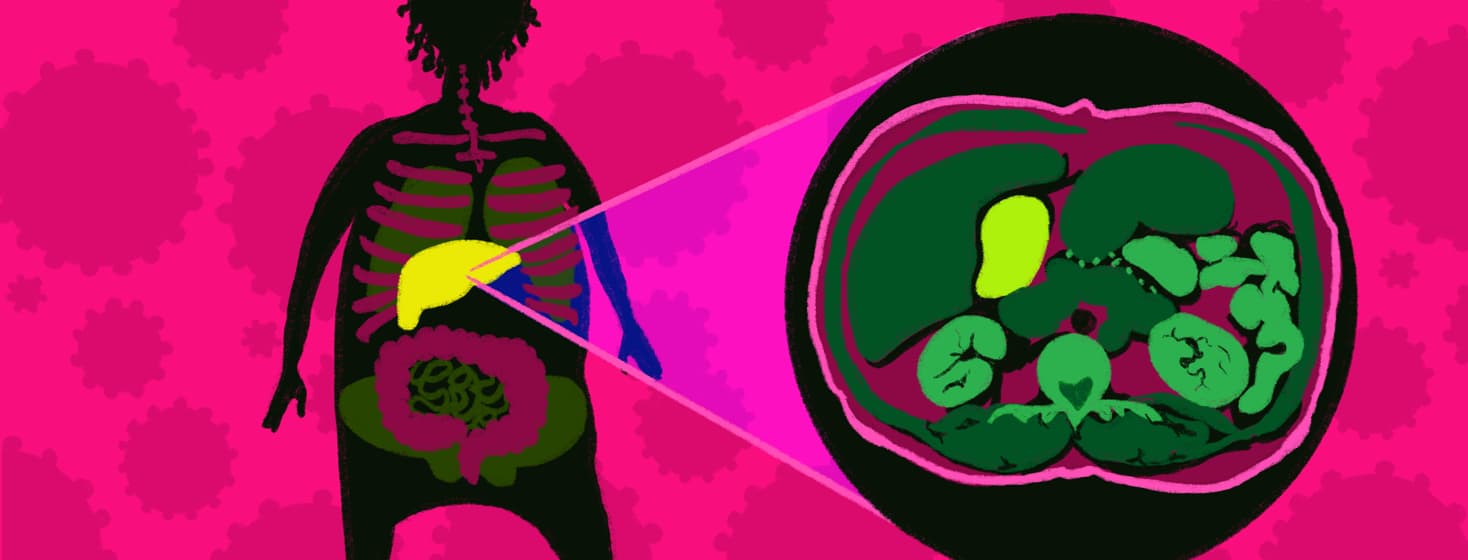
Imaging, or taking pictures of the inside of the body, is a part of treating and monitoring the hepatitis C virus (HCV). Imaging can be done to diagnose a condition or to track any changes it causes. Common types of body imaging are ultrasound, x-ray, CT (computed tomography) scans, and MRI (magnetic resonance imaging).
Imaging the liver can be done in a variety of ways and has many benefits when it comes to HCV.1
Liver damage and hepatitis C
HCV can cause damage to the liver over time. This damage can cause scarring. Once a person is diagnosed with HCV, their doctor will look for any signs of liver scarring or problems.1,2
Scarring that is reversible is called fibrosis. Scarring that is irreversible is called cirrhosis. Cirrhosis can lead to:1,2
- Liver failure
- Liver cancer
- Other serious health issues
Role of liver imaging in hepatitis C
There are blood tests that can be done to check how well the liver works. These tests look for different proteins made by the liver and other signs that point toward liver trouble. But these tests are not always the best predictors of liver damage.1-3
There are other problems with the body that can change the results of blood tests. It is also possible for results to not show how severe a liver issue actually is.1-3
In order to truly understand if the liver has scarring and how severe the scarring is, a person needs to have a liver biopsy. During a liver biopsy:1-4
- A needle is used to take a small sample of liver tissue (cells)
- The cells are looked at under a microscope
- Any scarring that is seen under the microscope can be scored
- Different scoring systems determine if a person has fibrosis or cirrhosis
But a liver biopsy is an invasive procedure. Along with the benefits, it has risks and side effects. Some types of liver imaging can be a good step to try before deciding that a biopsy is needed. Imaging can give a good understanding of liver damage. In some cases, imaging may be able to replace the need for a biopsy completely.1,3
Types of liver imaging
Each person’s experience with liver imaging may be different. But there are some types of imaging commonly used for people with HCV. These include:1-5Ultrasound: Ultrasound uses sound waves to create images of the inside of the body. The waves travel differently through water, solid tissue, organs, and bones. These differences make up the picture. Ultrasound images are not the clearest, but they can be made quickly. Ultrasound imaging is not painful.
Transient elastography (FibroScan): A type of ultrasound that is designed to measure the stiffness of the liver. The stiffer the liver, the more likely there is damage or scarring.
CT scan: CT scans use many different x-rays at once to create a 3-dimensional picture of the body. CT scans are clearer than ultrasounds. They also provide more detail than standard x-rays. But because they use x-rays, they expose the body to radiation. Radiation may have some risks.
Magnetic resonance elastography (MRE): MRE is a type of MRI scan. MRIs use magnetic fields to create images of the body.
Like ultrasound and FibroScan, MRE uses sound waves. MREs combine MRI technology and sound waves to determine how stiff the liver is.
MRI images are typically some of the clearest body images. But they can be expensive and cannot be used by people who have equipment like pacemakers.
They also require a person to sit or lie still for much longer than CTs or ultrasounds.
These are not all of the possible imaging options for HCV. Your doctor can help determine what tests you might need and how they can help in your situation.
Next steps and follow-up
Understanding if the liver is scarred and how bad scarring is can help you and your doctor make treatment decisions. It can also guide the next steps for follow-up.1
For example, cirrhosis increases the risk for liver cancer (hepatocellular carcinoma, HCC). Some people with cirrhosis may need to undergo regular screening.
Screening for liver cancer may involve both blood tests and imaging. Your doctor will work with you to determine your specific follow-up plan.1,6

Join the conversation